


このカタログについて
| ドキュメント名 | 【資料】CAR-T開発のための高性能ソリューション(英語版) |
|---|---|
| ドキュメント種別 | その他 |
| ファイルサイズ | 1Mb |
| 取り扱い企業 | ザルトリウス・ジャパン株式会社 (この企業の取り扱いカタログ一覧) |
この企業の関連カタログ


このカタログの内容
Page1
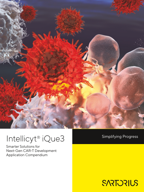
Intellicyt® iQue3
Smarter Solutions for
Next-Gen CAR-T Development
Application Compendium
Page2
Contents
Faster, Smarter Flow Cytometry to
Advance CAR-T Discovery and Development
Introduction .................................................................................................................................................................3
Rapid, Microvolume, High-Throughput Sampling Technology ........................................................3
Powerful Multiplexing Approach Through Encoding Technology .................................................3
Integrated Software Solution ..............................................................................................................................4
Conclusion: Reduce Errors and Time to Actionable Results .............................................................4
Applications of the iQue® Advanced
Flow Cytometry Platform
High-Throughput Cytokine Profiling in Human Peripheral Blood Mononuclear Cells .........4
Screening Ex Vivo Conditions Which Increase Memory T Cell Frequency ...............................7
Multiplex T Cell Activation Assay for Phenotypic Screening of Kinase Inhibitors ................... 9
Find out more
For more information, please visit
www.sartorius.com/car-t-research
Page3
Faster, Smarter Flow Cytometry to
Advance CAR-T Discovery and Development
Introduction Rapid, Microvolume,
High-Throughput Sampling
Technology
Chimeric antigen receptor T cell (CAR-T)-based therapy, The iQue3 advanced flow cytometry platform has rapid,
using engineered T cells to fight cancer, is a transfor- microvolume, high-throughput sampling technology
mative technology like gene therapy or regenerative based on flow cytometry that delivers rich content with
medicine that may forever change the landscape of sampling times of 5 minutes for a 96-well plate and
medicine. CAR-T cell-based therapy uses immune under 20 minutes for a 384-well plate. An entire plate
cells — either the patient’s own (autologous) or from a of data is processed at one time because samples are
donor (allogeneic) — that have been genetically trans- delivered to detectors in an air-gap delimited stream.
formed and are then infused into the patient to bind to The technology requires less than 10 µL, preserving
cancer cells and destroy them. precious sample for downstream analysis while reducing
reagent costs.
The discovery and development process for CAR-T
therapies has many steps, including:
Powerful Multiplexing Approach
Isolation of circulating T cells Through Encoding Technology
Early in vitro assays deploying specific tumor cell models
and understanding immune cell activation The iQue advanced flow cytometry platform performs
Imaging and flow cytometry to assess biological high-throughput assays with cells and beads in sus-
responses pension, enabling a powerful multiplexing approach
through encoding technology. Encoding technology
This application compendium demonstrates how allows screening against multiple target antigens in the
you can advance and accelerate your CAR-T-based same experiment because multiple populations of cells
discovery by illustrating some of the applications of the or beads bearing different antigens of interest can be
Intellicyt® iQue3 advanced flow cytometry platform. combined into the wells of assay plates.
This ability to simultaneously assess parameters
from multiple cell types in a single well makes the
iQue platform ideal for researchers delving into the
complexities of the immune system, such as changes
in signaling molecules and cell function. For example, it
allows measurement of T cell activation by proliferation,
cytokine secretion, and changes in subtype populations
(immunophenotyping) in the same analysis, for deeper,
more nuanced understanding of the biology.
3
Page4
Integrated Software Solution Conclusion: Reduce Errors and
Time to Actionable Results
The iQue advanced flow cytometry platform includes
Forecyt® Software to produce actionable data by To perform an effective screening campaign, the
generating plate heat maps, histograms, plots, dose screening technologies and reagents you use must have
response curves, and profile maps. A multiplate analysis the speed and sensitivity to support high-throughput,
feature, called Panorama, generates an analytical “big cost effective analysis, and meaningful data. The iQue
picture” that automatically compares, identifies, and advanced flow cytometry platform allows you to gain
ranks data across multiple plates of an experiment, as deeper insights with multiple parameter data acquisition
well as multiple plates in an experiment over several for multiple cell types, expand the scope and scale of
days. In addition, criteria threshold slider bars adjust data your research with fast, high-throughput flow cytometry
on the fly for real-time “what if” analyses of plate data capabilities, save on reagent costs and conserve your
with the click of a mouse. limited samples, and streamline your workflows with
powerful, built-in data analysis and visualization tools.
Forecyt Software is purpose-built to manage large data
sets from microtiter plates, and includes data analysis
and visualization tools that let you work with your data
in real time instead of waiting for batch analysis tasks.
Immediately see the impact of your actions for better,
more efficient insight.
Applications of the iQue®
Advanced Flow Cytometry Platform
High-Throughput Cytokine Profiling in
Human Peripheral Blood Mononuclear Cells
Immune function involves a coordinated set of secreted the ability to multiplex the detection of multiple proteins
protein signals across multiple cell types, and the regu- simultaneously; however, these assay protocols are often
lation of such signals depends on cytokine-dependent long and laborious. In addition, many bead-based assays
cell-to-cell communication. These complex signaling require multiple wash steps, which limits the ability to
pathways between cells are an important target across automate the assay and causes bead loss, increasing
the drug discovery process, from primary screening to variability in the results.
toxicity profiling.
The Multicyt® Qbeads Plexscreen reagent leverages the
Profiling of secreted proteins is ideally achieved by benefits of the iQue advanced flow cytometry platform
the simultaneous detection of multiple proteins. This to provide a unique, no-wash protocol that is ideal for
maximizes the contextual and correlative value of the medium- to high-throughput screening (Figure 1).
data. Traditional technologies, such as plate-based en- Qbeads are assembled into user-specified multiplate kits
zyme-linked immunosorbent assays (ELISAs), are limited that can contain up to 30 analytes (Figure 2). Compared
because they are inherently single-endpoint readouts. to other bead-based protein detection assays, Qbeads
Newer technologies, such as bead-based ELISAs, offer provide an easier screening workflow, lower cost, faster
4
Page5
Qbeads-figure1
Qbeads Assay Principles
A. B. Free soluble C.
protein Complete
Qbead sandwich complex
Fluorescently
labeled
Protein-specific Captured detection
capture antibody protein antibody
Figure 1: Qbeads function by performing a sandwich ELISA on the surface of beads. (A) Qbeads are coated with analyte-specific capture
antibodies. (B) Qbeads capture analyte that binds to the analyte-specific antibodies on the bead surface. (C) A fluorescently-labeled
detection antibody is added, causing the beads to fluoresce in proportion to the analyte concentration in the sample and providing a signal
detectable on an iQue platform.
Qbeads-figure2
Multiplexing Qbeads
A. B.
RL 1 Color Bead 1
Bead 2
RL 2 Color
Bead 3
RL1
Figure 2: The simultaneous detection of multiple analytes is possible by using mixtures of beads specific for different analytes. (A) Beads
1, 2, and 3 are each encoded with unique intensities of two fluorescent colors, RL 1 and RL 2. Each bead is specific for one type of analyte,
represented by the different shapes. (B) After the beads are run through the iQue platform, they are categorized by their fluorescence
signature, which allows the binding signals for the appropriate analytes to be separated in a multiplexed sample.
plate read times, and lower sample volume requirements. Methods
These advantages allow the practical screening of large On three consecutive days, cryopreserved PBMCs
libraries for the ability to modulate secreted protein from a single donor were thawed and treated in 384-
profiles. well plates with known T cell stimulating agents. The
T cell stimulation methods included: (1) co-treatment
Here, we present a case study using Qbeads to profile with CD3 and CD28 antibodies (top dose = 5 μg/mL for
cytokine secretion in human peripheral blood mononu- each antibody, (2) co-treatment with PMA (top dose =
clear cells (PBMCs). We performed a multi-day study to 10 ng/mL) and ionomycin (top dose = 10 μg/mL), and
examine cytokine secretion in a 7-plex assay representing (3) treatment with Phytohemagglutinin (PHA; top dose
activation of Th1/Th2/Th17 T helper cells. = 5 μg/mL). The secretion of IL-2, IL-4, IL-6, IL-10, IL-17A,
IFNγ, and TNFα were detected and quantified directly
Our results highlight the turnkey solution represented by from the supernatants.
the Qbeads reagents on the iQue platform, and illumi-
nate the tremendous value of multiplexing, not only in
time and cost savings, but also in the ability to generate
correlative data in a physiologically relevant context.
5
RL2
Page6
Results Assay miniaturization possible on the iQue platform
We observed different cytokine secretion profiles allowed a cost effective screen of a dose response
depending on which PBMC stimulation method we for PBMC stimulation, and revealed PHA as a weak
used. A subset of the cytokines assayed (IL-4, IL-10, IFNγ inducer rather than a negative treatment, thus
and IFNγ) highlights some of the most significant eliminating the need for time-consuming dose-finding
differences between the three stimulation methods. experiments.
PMA/Ionomycin stimulation caused no observable IL-4
or IL-10 secretion at any dose but induced a strong IFNγ The Multicyt Qbeads portfolio presents an opportunity
response (Figure 3). CD3/CD28 and PHA produced to elevate the screening of secreted proteins to an
a similar cytokine profile — IL-4 is negative at all doses unprecedented level of efficiency by offering a stream-
while both IL-10 and IFNγ are positive. lined, multiplexed workflow at significantly lower cost
than many existing technologies.
Summary
IFNγ secretion across the three stimulation methods References
emphasizes the value of screening in a dose response Narang, R., et al. Application of Multicyt Qbeads for
series. Of the three stimulation methods, CD3/CD28 was High Throughput Cytokine Profiling in Human Periph-
the most potent IFNγ inducer, followed by PMA/Iono- eral Blood Mononuclear Cells. Sartorius Application
mycin, then PHA. Interestingly, if we had screened PHA Note, 2014.
in a single-dose format, with a dose in the middle of the
range tested here, it would have likely scored negative
for IFNγ secretion.
PMA/Ionomycin CD3/CD28 PHA
40000
30000
IL-4 20000
MFI
10000
Limit of Detection
40000
30000
IL-10
MFI 20000
10000
Limit of Detection
0
300000
200000
IFNγ
MFI
100000
Limit of Detection
0
Dose Dose Dose
Figure 3: Using Qbeads shows different cytokine profiles for three different PBMC stimulation methods. The dotted line represents
3 standard deviations of the mean response for unstimulated controls (N=96). All dose response series are 1:2 serial titrations.
6
Day 1 Cytokine Profiles for 3 Different PBMC Stimulation Methods
Page7
Screening Ex Vivo Conditions Which
Increase Memory T Cell Frequency
The ex vivo expansion of T cells is a critical process in Methods
the biomanufacturing of adoptive cell therapies, such as We performed screening studies using human
chimeric antigen receptor (CAR) T and tumor infiltrating peripheral blood mononuclear cells (PBMCs) from
lymphocyte (TIL) therapies. Recent clinical studies show three donors cultured in a single, 96-well plate over
that a correlation between the persistence of subsets of three days and using 14 different culture conditions.
functional memory T cells, including T memory stem cells In each assay well, we distinguished live immune
(Tscm), central memory T cells (Tcm) and other less differen- cells from dead cells by staining with a fluorescent
tiated T cell subsets, is responsible for long term anti-tumor membrane integrity dye that only enters dead cells or
responses in patient outcomes. This suggests ex vivo T cell those with a compromised membrane and staining
expansion protocols generating higher percentages of the nucleic DNA by intercalation.
Tscm and Tcm in the T cell product are critical to significant
clinical improvements in adoptive cell therapies. We immunophenotyped live cells by staining with a
fluorescent antibody panel to separate CD3+ (T cells),
Here, we show results from a robust T memory cell and CD3- (non-T cells), CD4+ (T helper cells) and CD8+
cytokine profiling assay optimized for 96- or 384-well (T cytotoxic cells). The antibody panel also included
microtiter plates that runs on the iQue advanced flow five different T cell surface markers for T naive/memory/
cytometry platform. The Intellicyt® T Cell Memory Cell effector cell phenotyping: CD45RA, CD45RO, CD27,
and Cytokine Profiling Assay Kit (TCA Kit) was developed CD62L, and CD95. Effector cytokines secreted by
to address the need to monitor T cell phenotype and ex vivo expanded T cells, including pro-inflammatory
function for improved ex vivo expansion protocols where cytokine IFNγ and anti-inflammatory cytokine IL-10,
profiling of memory subsets is crucial. This assay includes were measured in a sandwich immune assay format by
antibody markers to identify T cells and identifies naive, two different Qbeads, included in the same well (see
Tscm, Tcm and TEFF. In addition, a functional analysis Figure 4). We analyzed T cell expansion time course data
of the cells is performed by quantitating the levels of and produced the series using the Forecyt software
secreted IFN and IL-10 simultaneously with phenotypic package.
measurements.
One Well Self-Renewal Potency
Cd3+ T Cells Cd4+ T Helper
TN TSCM TCM TTM TEM TEMRA TTE
Activation CD45RA: + + - - - + +
CD45RO: - - + + + + -
CD27: + + + + - - -
Cd8+ T Cytotoxic CD62L: + + + - - - -
CD95: - + + + + + +
Cytokine Secretion
Live
Dead Pro-Inflammatory Anti-Inflammatory
Cytokine IFNγ Cytokine IL-10
Cell Membrane
Integrity Dye
IFNγ IL-10
Capture Capture
Bead Bead
Cytokine Detection On Qbeads®
Figure 4: Schematic of this multiplex assay design. Multiplexed measurement in each well.
7
Page8
Results increased in the wells treated with a cocktail containing
Figure 5 shows the results from Donor 1 on Day 5. Tcm IFNß compared to the well treated with CD3/CD28
(especially cytotoxic CD8+ Tcm) increased in frequency Dynabeads alone. CD3+ T cell counts in culture supple-
in six cocktails containing IFNß, and Tscm (particularly mented with cytokine cocktails containing IFNß also
helper CD4+ Tscm) increased in frequency increased in slowed down cell proliferation by approximately 50%
four cocktails containing IL-21 (Figure 5A). In Figure 5B, (Figure 5C).
the 2D plots (CD27 vs. CD62L) show Tcm cell frequency
A. B.
Tcm Tscm Tcm Comparison
Donor 1 Donor 1 Treatment 2 Treatment 7
5.0 5.0
(CD3/CD28 (CD3/CD28Dynabeads
IL-21 Dynabeads only) + IL4/IL7/IFNβ)
4.0 4.0
BO1 & RA-RO+CD95+ GO1 & RA-RO+CD95+
3.0 3.0 2 9 11 14 107 107
106 Tcm 106 Tcm
2.0 2.0 105 105
2 104 104
1.0 1.0 -105 Tem Ttm -105 Tem Ttm
0.0 0.0 -104 -104
0 2 4 6 8 10 12 14 16 0 2 4 6 8 10 12 14 16 -104 -105 104 105 107 -104 -105 104 105 107
16 Treatments 16 Treatments CD27 (RL2-H) CD27 (RL2-H)
Donor 1 Donor 1
5.0 5.0 C.
IFNβ
4.0 4.0 CD3+ T Cell Count
7 10 12 13 15 16
3.0 3.0 Donor 1
2.0
2.0 2.0 1.8
2 2 1.6
1.0 1.0 1.4
1.2
6 9 11 14
0.0 0.0 1.0
0 2 4 6 8 10 12 14 16 0 2 4 6 8 10 12 14 16 0.8
16 Treatments 16 Treatments 0.6
0.4
0.2 7 10 12 13 15 6
1 2 3 4 5 6 7 8 9 10 11 12 13 14 15 16 0.0
0 2 4 6 8 10 12 14 16
CD3|CD28 - + + + + + 16 Treatments
Dynabeads
IL-4 | IL-7 - - + + + +
IL-6 - - - + - - - + + + - - - + + -
IL-15 - - - - + - - + - - + + - + + +
IL-21 - - - - - + - - + - + - + + - +
IFNβ - - - - - - + - - + - + + - + +
1 2 3 4 5 6 7 8 9 10 11 12 13 14 15 16
CD3|CD28 - + + + + +
Dynabeads
IL-4 | IL-7 - - + + + +
IL-6 - - - + - - - + + + - - - + + -
IL-15 - - - - + - - + - - + + - + + +
IL-21 - - - - - + - - + - + - + + - +
IFNβ - - - - - - + - - + - + + - + +
Figure 5: Increased T memory cell frequency after ex vivo expansion in cytokine supplemented media. Cytokine cocktails containing IFNβ or
IL-21 increase the T central memory cell frequency or T stem cell-like memory cell frequency, respectively. Example of Donor 1 on Day 5 was
shown here. (A) T central memory cells (Tcm), especially CD8+ Tcm showed frequency increase in 6 cocktails containing IFNβ; T stem cell-like
memory cells (Tscm), especially CD4+ Tscm showed frequency increase in 4 cocktails containing IL-21. (B) The well-scan of 2D plots (CD27 vs.
CD62L) showed Tcm cell frequency increase in the well treated with a cocktail containing with IFNβ, compared with the well treated with CD3 |
CD28 Dynabeads. (C) CD3+ T cell counts showed cocktails with IFNβ slowed down the cell proliferation about 50%.
8
CD8+ CD4+
CD8 Tcm (Folds of Donor 1_CD3/CD28) CD4 Tcm (Folds of Donor 1_CD3/CD28)
CD8 Tscm (Folds of Donor 1_CD3/28) CD4 Tscm (Folds of Donor 1_CD3/28)
CD3 Cell Count (Folds of CD62L (BL1-H)
Donor 1_CD3/28)
CD62L (BL1-H)
Page9
Summary Multiplexed cell and secreted cytokine measurements
Here we showed that media supplements had in a single assay offers an improvement over common
profound effects on the final T memory subset, immunology research workflows that require multiple
cellular composition, and cytokine release. Despite assays run on different platforms. The kit allows spatial
the complexity of T cell biology, the simple assay and temporal analysis of T memory cell phenotypes
and analysis workflow of the Intellicyt® Human T and functions at different stages, all in a single high-
Cell Memory and Cytokine Profiling Kit could be content, miniaturized assay that saves precious samples,
useful for adoptive T cell therapy workflows. The kit decreases reagent costs, and enhances data integrity.
is designed for ease-of-use by combining cells and
beads, which allows multiplexing measurements References
from each assay well. Liu, Z., et al. An Optimized, Multiplexed Assay for
Screening Ex Vivo Conditions which Increase Memory
T Cell Frequency. Sartorius Application Note, 2019.
Multiplex T Cell Activation Assay for
Phenotypic Screening of Kinase Inhibitors
Activation of the T cell receptor (TCR) pathway in naive peripheral blood mononuclear cells (PBMCs) stimulated
and effector T cells leads to T cell activation, proliferation, with anti-CD3/CD28 beads. We acquired samples on
and cytokine production. Modulating TCR engagement the iQue platform and assessed cell proliferation in
and signaling pathways using biologics, small molecules, viable CD4 and CD8 lymphocytes, as well as early/late
or genetic engineering is highly relevant to many thera- activation markers CD69, CD25 and HLA-DR. To assess
peutic areas, including cancer immunotherapy, adoptive T cell function, we quantified the levels of secreted IFNγ
cell therapy, and vaccine development. and TNFα. We analyzed data and generated heat maps,
IC50 curves, and cytokine levels using Forecyt software.
Perturbations leading to increased hyper-responsive We used Profile Maps, a unique analysis tool of Forecyt
TCR signaling and enhanced T cell activation is a to integrate assay metrics with Boolean logic to quickly
major cause of autoimmune disease. Genetic defects, locate hits using defined multiplexed criteria.
mutations, and other mechanisms resulting in increased
T cell kinase activity are involved in many autoimmune Results
pathologies, making them attractive targets for the The TCA kit can generate data on 15 different param-
direct inhibition of T cell activation. eters, but as an example, Figure 6 demonstrates a
plate-level analysis showing the percentage of viable
The development of drugs and therapies regulating CD4 T cells that express the early activation marker
TCR activity requires assays to profile T cell function and CD69 from Plate 1. Using this visualization tool, we can
health. Here, we report on an optimized, high-content, quickly identify compounds that have inhibited expres-
multiplexed assay using high-throughput flow cytometry sion of CD69 (CD69+ cells are in the rectangular gate),
to measure T cell activation. as well as KI that drastically reduced CD4+ T cell viability
(wells with no cells, for instance, well H2).
These studies demonstrate the insight provided by the
use of the iQue advanced flow cytometry platform for The plate-level view shows examples of inhibitors that
phenotypic screening of small molecules affecting T cell affect different kinase families. In wells containing media
activation. alone, 86% of the CD4 cells are activated, as assessed by
CD69 expression and treatment with the FDA-approved
Methods Jak 1/2 inhibitor Ruxolitinib, which reduced the percent-
We used the Intellicyt® Human T Cell Activation Cell age of CD4+CD69+ cells down to 55%. The Src inhibitor
and Cytokine Profiling Kit (TCA Kit) to screen a 152 PP2 dramatically inhibited CD69 expression as only 3%
small molecule library of kinase inhibitors (KI) for their of the CD4 T cells expressed CD69. Other compounds
ability to inhibit human primary T cell activation in showed a range of CD69 inhibition.
9
Page10
1 2 3 4 5 6 7 8 9 10 11 12
A
B
C
D
E
F
G
H
Negative Positive
Side Scatter
(SSC-H)
Well-A1 Well-A4 Well-B5 Well-B10 Well-D7 Well-E10
A01 and CD A04 and CD4+ B05 and CD4+ B10 and CD4+ D07 and CD4+ E10 and CD4+
Media only Doramapimod AS-703026 Ruxolitinib JAK 1/2 PP2 Src Kinase KN-93 CaMKII
p38MAPK MARK/ERK Kinase
07 07 07 07 07 07
CD4+CD69+ CD4+CD69+ CD4+CD69+ CD4+CD69+ CD4+CD69+ CD4+CD69+
05 85.54% 05 52.53% 05 37.3% 05 54.87% 05 3.26% 05 67.09%
04 04 04 04 04 04
04 04 04 04 04 04
03 03 03 03 03
-104 -103 104 105 106 107 -104 -103 104 105 106 107 -104 -103 104 105 106 107 -104 -103 104 105 106 107 -104 -103 104 105 106 107 03
-104 -103 104 105 106 107
CD69
Figure 6: Compounds that inhibit CD69 expression. A plate-level view (from Plate 1) of CD69 expression in T helper cells generated
in Forecyt software. The CD69 expressing cells are found in the rectangular gate. Wells highlighted in red are shown below with the
indicated KI and the % of CD69 CD4+ cells found in the inset of each dot blot.
To integrate the data for all 15 activation metrics, and cytokine secretion (wells highlighted in yellow in
we used the Forecyt Profile Map data tool in the Figure 7). The overlay line graph in Figure 7 ranks all
Panorama feature (see Figure 7). The left panel of the hits and the level of inhibition, providing easy
shows the 11 metrics from this screening study and visualization for each metric. For example, treatment
the user-defined threshold for each of these. Using with some KI completely inhibits all activation metrics,
the Profile Map, we identified 27 different KI that whereas other compounds have a greater impact on
inhibited expression of all T cell activation markers specific phenotypic markers or cytokines.
10
Page11
A. B.
Criteria Plate 1 Plate 2
INFγ+ (pg/mL) <= 1,000.00
TNFα (pg/mL) <= 1,000.00
% live cells >= 40.0
CD4+CD69+ as % of CD4+ <= 70.0
CD8+CD69+ as % of CD8+ <= 70.0 C.
Hits Ranking
100
CD4+CD25+ as % of CD4+ <= 70.0
90
80
CD8+CD25+ as % of CD8+ <= 70.0 70
60
CD4+HLA-DR+ as % of CD4+ <= 30.0 50
40
30
CD8+HLA-DR+ as % of CD8+ <= 30.0
20
10
Count of CD4+ >= 140.0 0
Count of CD8+ >= 140.0
Plate ID and Well ID
Figure 7: Data generated using the unique Profile Map function of Forecyt Software. To identify hits that inhibited all T cell activation metric,
we generated Profile Maps using the Panorama feature in Forecyt. The user-defined threshold level of each of the desired metrics is shown in
(A), while the specific hits from the 2 plates are shaded in yellow (B). We generated a line graph ranking the hits based on the % of CD69 CD4+
cells (red line) and overlaid line graphs for 5 additional metrics for each of the hits (C). These visualization tools provide easy insight into how the
various KIs impact different T cell activation metrics.
Summary References
The Intellicyt® TCA kit can be used as a primary or Liu. Z., et al. A kinase inhibitor phenotypic screen using
secondary phenotypic screen in both the biologics and a novel multiplex T cell activation assay. Sartorius
small molecule workflows. Additionally, the TCA kit is Application Note, 2019.
used for functional studies in the immuno-oncology
space and for characterizing T cells during cell manu-
facturing processes. The TCA kit is applicable to many
functional assay workflows including development of
checkpoint inhibitors and cell therapies and during cell
manufacturing.
11
Percentage (%)
Plate 1-H08
Plate 1-B08
Plate 2-B08
Plate 1-C04
Plate 2-E11
Plate 1-D07
Plate 1-D09
Plate 2-G07
Plate 1-D10
Plate 1-E08
Plate 1-B05
Plate 1-G05
Plate 1-E07
Plate 2-G11
Plate 1-G06
Plate 1-B04
Plate 1-A03
Plate 1-A04
Plate 1-A10
Plate 1-B10
Plate 1-C03
Plate 1-F10
Plate 1-E10
Plate 2-B10
Plate 1-D11
Page12
Sales and Service
Contacts
For further contacts, visit
www.sartorius.com
Essen BioScience, A Sartorius Company
www.sartorius.com/intellicyt
info.intellicyt@sartorius.com
North America APAC
Essen BioScience Inc. Essen BioScience K.K.
300 West Morgan Road 4th Floor Daiwa Shinagawa North Bldg.
Ann Arbor, Michigan, 48108 1-8-11 Kita-Shinagawa
USA Shinagawa-ku, Tokyo
Telephone +1 734 769 1600 140-0001
Japan
Europe Telephone +81 3 6478 5202
Essen BioScience Ltd.
Units 2 & 3 The Quadrant
Newark Close
Royston Hertfordshire
SG8 5HL
United Kingdom
Telephone +44 (0) 1763 227400
Learn More About Sartorius
Specifications subject to change without notice.
©️ 2020, Essen BioScience, Inc., part of the Sartorius Group. All Rights Comprehensive Solutions for CAR-T
Reserved. Intellicyt, iQue, iQue3, iQue Screener PLUS, Forecyt, Multicyt, and
all names of Intellicyt products are registered trademarks and the property
of Essen BioScience unless otherwise specified. Intellicyt is a Sartorius brand. Discovery and Development at
Printed in the EU or US on paper bleached without chlorine.
Version 1 | 2020 | 04 www.sartorius.com/car-t-research.